The guide · Mobility, Early recovery
Why won't my knee straighten after ACL surgery?
Extension loss is what rehabilitation protocols watch most closely in the early weeks. What causes it, why it is treated as urgent, and what is usually done.
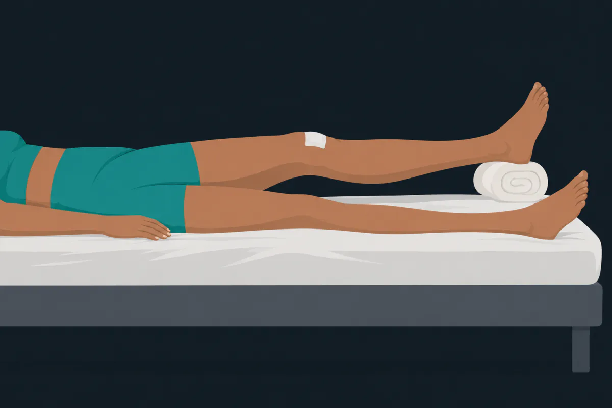
A knee that will not go flat is one of the first things people notice after an ACL reconstruction, and it is the thing physiotherapists watch most closely. You lie down, prop the heel on a rolled towel, and there is a gap under the back of the knee that the other leg does not have. This article covers what causes that gap, why protocols treat it as the early priority rather than something to catch up on later, and what is usually done about it.
Your surgeon’s and physiotherapist’s instructions come first, including how hard extension should be worked and which exercises suit your graft and any repair done at the same time. Nothing here replaces them.
What “straight” actually means
Extension is the straightening direction, measured in degrees away from straight. A knee resting at zero is flat. A knee that lacks ten degrees of extension sits ten degrees short of flat and will not press down onto the bed.
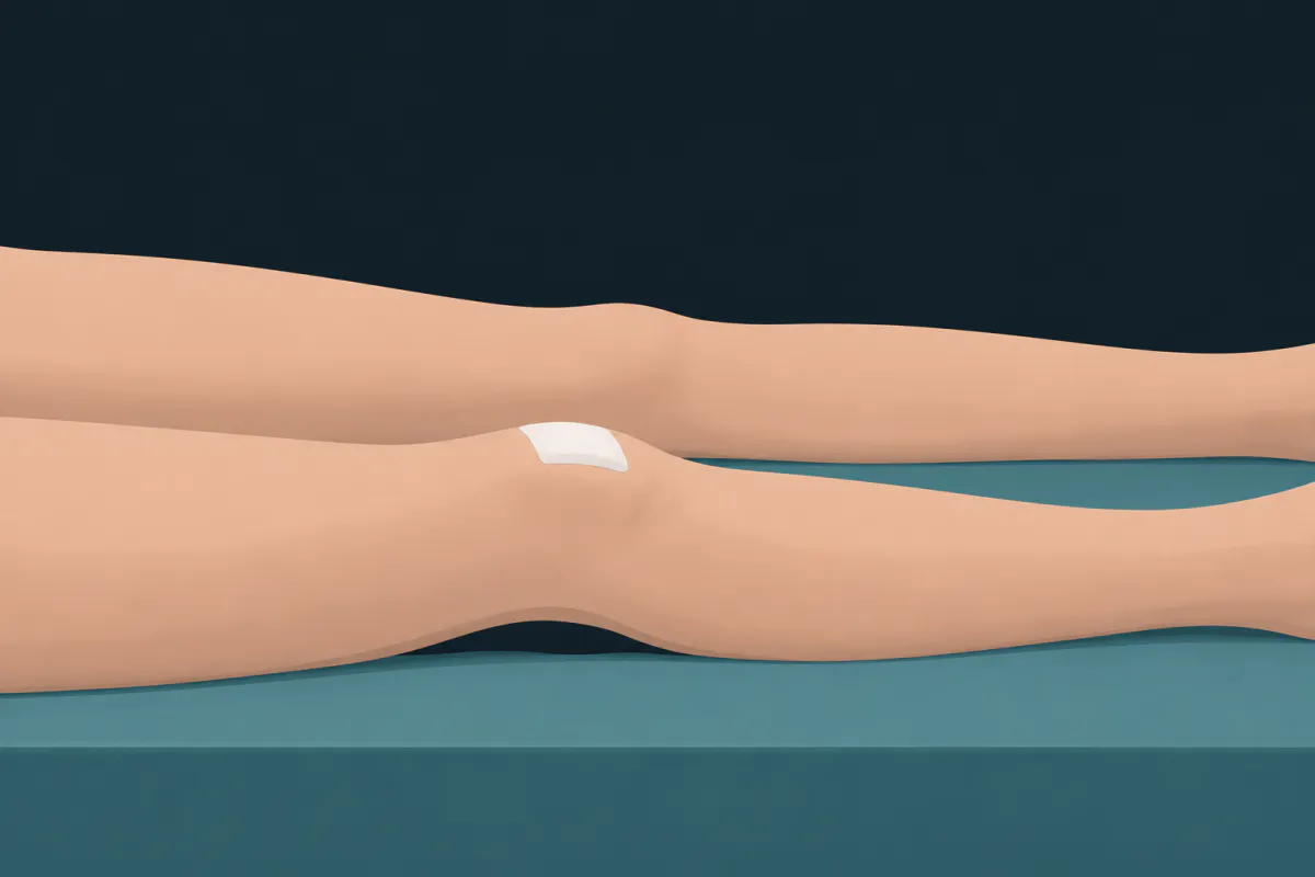
The target, though, is not zero. Plenty of knees go past flat into a few degrees of hyperextension, and that is normal anatomy rather than looseness. The comparison that matters is your other leg. When Shelbourne and Gray followed patients for a minimum of ten years after reconstruction, they defined normal knee motion as extension within two degrees of the uninvolved knee, hyperextension included, and flexion within five degrees. That is a tight definition, and it is deliberately tight: the uninjured side is the only fair reference you have.
This is also why extension gets more attention than flexion, even though flexion is the loss people notice first. A knee that cannot bend far enough makes stairs and car seats awkward, and it tends to improve as swelling settles. A knee that cannot straighten changes how you stand and walk, and the research has treated it as the more consequential of the two.
Why the knee resists straightening in the early weeks
Several ordinary things push in the same direction at once.
- Swelling. Fluid inside the joint raises the pressure in it, and a pressurised joint is most comfortable slightly bent. That is the position the knee drifts into whenever you stop thinking about it.
- Pain and guarding. The hamstrings behind the thigh tighten protectively, holding the knee a few degrees short.
- The quadriceps not firing. Straightening is not only a passive matter of the joint allowing it. The thigh muscle actively holds the knee straight in standing and at the end of each step, and after surgery it is often slow to switch on.
- Position and habit. A pillow under the knee is the most comfortable arrangement available in the first fortnight, and it holds the joint bent for hours at a time. Sitting with the knee flexed and walking with a short, bent-knee gait do the same thing in smaller doses all day.
None of those are complications. They are the normal early state of a knee that has had bone drilled and a graft passed through it, and this is the reason most protocols say something about how to rest the leg rather than leaving it to comfort.
Why protocols treat extension as urgent
The consensus rehabilitation guideline from van Melick and colleagues puts regaining full extension among the goals of the earliest postoperative phase, alongside controlling swelling and pain, restoring quadriceps control and normalising walking. Noyes’ Knee Disorders sets out the reasoning that most protocols follow: extension is protected from the first days rather than recovered later, because a knee allowed to settle short of straight becomes considerably harder to straighten afterwards than a stiff knee is to bend.
The long-term argument comes from Shelbourne and Gray’s follow-up above. Their conclusion, reflected in the paper’s title, was that loss of normal knee motion compounds the other factors already known to contribute to arthritis after this surgery, such as meniscus and cartilage damage. It was a single-centre series of patellar tendon reconstructions, so it describes one surgeon’s population rather than every knee, and it cannot show that motion loss alone causes arthritis. It does explain why a few degrees that feel trivial at week three are treated seriously by people who have watched knees for a decade.
What is usually done about it
Early management is unglamorous and mostly about position and repetition: supporting the leg under the heel rather than under the knee so the joint hangs straight, controlling swelling, and getting the quadriceps contracting so the knee is actively held out at the end of each step. Extension work of some kind appears in almost every protocol in the first weeks, and the specific exercises, how long they are held and how firmly they are pushed are set by your physiotherapist.
For most people, that is enough. Noyes and colleagues ran a prospective study of 443 knees under a programme built around immediate motion and early intervention. At follow-up, 98% had a normal range of motion, and seven knees had a mild five-degree extension loss. Twenty-three knees, about 5%, needed something more than the standard programme: extension casts, gentle manipulation under anaesthetic, arthroscopic debridement, or a period of inpatient therapy under epidural anaesthetic. All twenty-three regained full motion, and no knee in the series ended with permanent arthrofibrosis.
Read that number both ways. Early motion works for the large majority, and needing extra help is neither rare nor a sign that the recovery has gone wrong. It is also why physiotherapists check extension at every appointment: the point of measuring weekly is to catch a knee that is not moving in the right direction while intervention is still simple.
When a block is not just stiffness
If extension stays blocked and the end of the movement feels like a firm stop rather than tightness, a cyclops lesion is one of the things that gets considered. It is a nodule of scar tissue in the notch at the front of the knee, sitting in front of the graft, first described on MRI by Recht and colleagues in patients whose knees would not fully straighten; the name comes from how it looks through the arthroscope.
Finding one on a scan is not the same as finding the explanation. In a European Radiology study of 113 patients scanned before surgery and at six months, one year and two years afterwards, cyclops lesions were present in about a quarter of knees, developed within the first six months, and were not associated with worse clinical outcomes at two years, whether by their presence or their size. Symptoms, examination and imaging have to agree before the lesion is blamed, and that judgement belongs to your surgeon.
Separately, some symptoms are not part of the expected picture at all. The NHS recovery guidance lists the standard warning signs after this surgery: a calf that is hot, red, swollen or painful, or new shortness of breath or chest pain, which can indicate a blood clot; spreading redness at the incision, discharge from the wound or a fever; new weakness or numbness in the foot; and a knee that locks and cannot be moved. Any of those means contacting your surgical team, your GP or an urgent care service the same day.
Tracking extension between appointments
If you use ACL Recovery Tracker, extension has its own field in the mobility log, entered by hand or measured with the phone’s gyroscope, and you can record a baseline from your uninjured leg to compare against. Logging both sides gives you the comparison the research uses instead of a memory of how flat the knee looked last week, and the trend view is a more useful thing to bring to an appointment than a single reading. It is a free ACL tracker app for iOS and Android, and you can use it next to whatever stretching plan your physio has set.
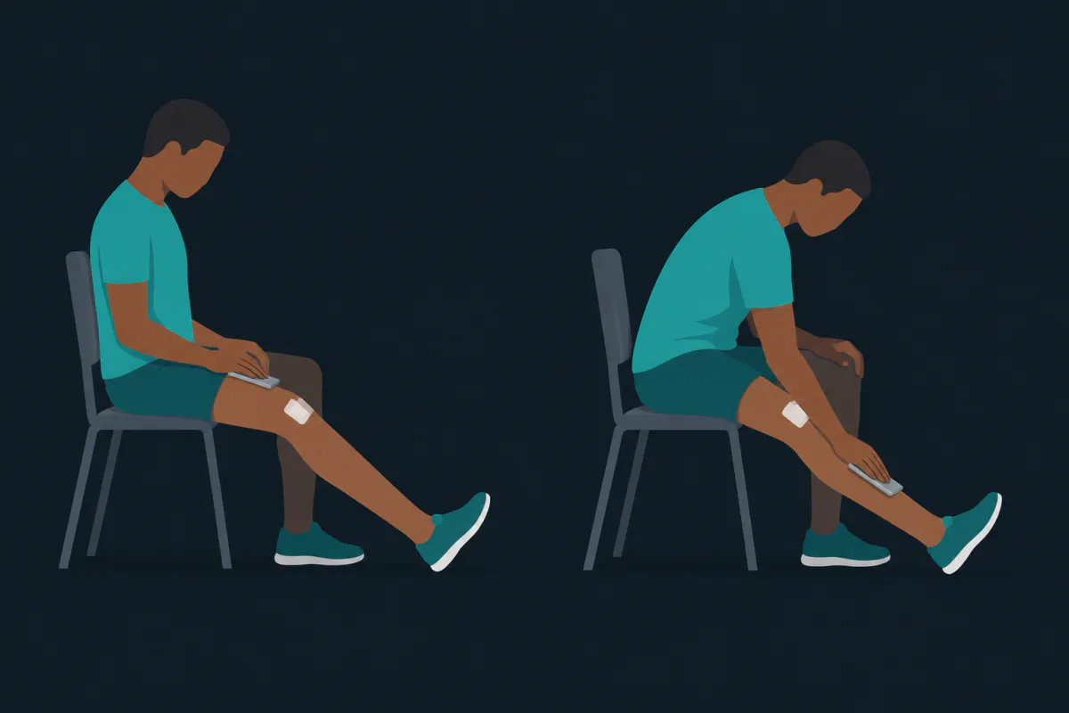
A gap under the knee at week two is ordinary, and most of them close with the plain early work. What is worth doing is noticing it, measuring it rather than guessing, and saying so at your next appointment if it is not changing.
The illustrations in this article were generated with AI.
Sources
- van Melick N, et al. Evidence-based clinical practice update: practice guidelines for anterior cruciate ligament rehabilitation based on a systematic review and multidisciplinary consensus. Br J Sports Med. 2016;50:1506-1515.
- Noyes FR, Berrios-Torres S, Barber-Westin SD, Heckmann TP. Prevention of permanent arthrofibrosis after anterior cruciate ligament reconstruction alone or combined with associated procedures: a prospective study in 443 knees. Knee Surg Sports Traumatol Arthrosc. 2000;8(4):196-206.
- Shelbourne KD, Gray T. Minimum 10-year results after anterior cruciate ligament reconstruction: how the loss of normal knee motion compounds other factors related to the development of osteoarthritis after surgery. Am J Sports Med. 2009;37(3):471-480.
- Recht MP, Piraino DW, Cohen MAH, Parker RD, Bergfeld JA. Localized anterior arthrofibrosis (cyclops lesion) after reconstruction of the anterior cruciate ligament: MR imaging findings. AJR Am J Roentgenol. 1995;165(2):383-385.
- Cyclops lesions detected by MRI are frequent findings after ACL surgical reconstruction but do not impact clinical outcome over 2 years. Eur Radiol. 2017;27(8):3499-3508.
- Noyes FR, Barber-Westin SD, eds. Noyes' Knee Disorders: Surgery, Rehabilitation, Clinical Outcomes. 2nd ed. Elsevier; 2017.
- NHS. ACL (anterior cruciate ligament) surgery: recovering.