The guide · Mobility, Pain and swelling
Cyclops lesion after ACL surgery
What a cyclops lesion is, why a nodule of scar tissue can stop the knee straightening, how often it causes symptoms, and what is usually done about it.
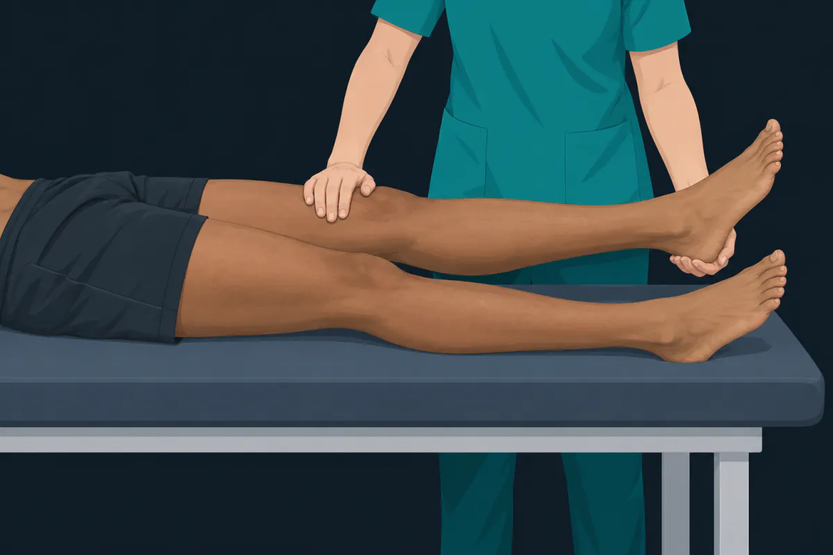
Most knees gain extension steadily in the first weeks after a reconstruction. A few stop short, and stay short, and in a small number of those the reason is mechanical: a lump of scar tissue sitting in the front of the joint, in the way of the last few degrees. That lump is a cyclops lesion, and this article covers what it is, how often it causes trouble, and what is usually done about it.
Whether your own knee’s extension is on track, and what to do if it is not, is a question for your surgeon and physiotherapist. Their assessment comes first; nothing here can substitute for someone putting their hands on the knee.
What a cyclops lesion is
The name dates from 1990, when Jackson and Schaefer described thirteen patients who, after an ACL reconstruction with a patellar tendon autograft, lost full extension and developed an audible, palpable clunk as the knee came towards straight. At arthroscopy each had a nodule of tissue sitting just in front of and to the side of the tibial tunnel — the hole drilled in the shin bone for the graft. The mass with its surface vessels looked, through the arthroscope, like a single eye, and the name stuck.
In more formal language it is localised anterior arthrofibrosis. Arthrofibrosis means scar tissue forming inside a joint; the generalised kind stiffens the whole knee in every direction, while a cyclops lesion is the focal version — one discrete nodule in the notch at the end of the thigh bone, in front of the graft. Aloisi and colleagues removed and examined this tissue from thirteen lesions found in a series of 126 knees and reported dense fibroelastic nodules. Scar tissue, in other words, organised into a lump. Notably, they were looking during primary reconstructions, so the tissue can already be there in a knee whose ACL is torn but not yet reconstructed.
Two things it is not. It is not the graft failing, and it is not a new tear. Mechanically it behaves like a doorstop: the knee runs out of room before it runs out of ligament.
How often it actually causes symptoms
There are two different numbers here, and confusing them causes needless worry. One counts lesions; the other counts problems.
For lesions, Facchetti and colleagues scanned 113 patients with a complete ACL tear before surgery and again afterwards, and found cyclops lesions on MRI in 25 per cent at six months, 27 per cent at one year and 33 per cent at two years (the later figures from 75 and 33 patients, as follow-up thinned). Their conclusion was that these lesions are frequent findings that did not affect scores on a standard knee questionnaire over two years. Seen on a scan, then, the tissue is common and usually silent.
For problems, the numbers are much smaller. Noailles and colleagues pooled ten studies in a systematic review and reported the incidence of symptomatic cyclops lesion ranging from 1.9 to 10.9 per cent — a spread that reflects how differently studies define and look for it. In a single large series, Delaloye and colleagues reviewed 3,633 primary reconstructions from the SANTI database and found 65 patients, 1.8 per cent, who had a second operation for cyclops syndrome. In the MERIscience cohort, Tarchichi and colleagues reported 197 patients treated surgically for it, which was 4.3 per cent of the primary reconstructions over that period.
So scar tissue in this spot is a common incidental finding, while a blocked knee that needs treating happens to a small minority, in the low single figures in most series. “Cyclops lesion” on a scan report is not by itself a diagnosis; what makes it one is the knee.
What it feels like
The picture that sends people looking this up is usually some combination of:
- the last few degrees of straightening not coming, and not coming even when someone else moves the leg
- a clunk or thud as the knee approaches full extension, which was the feature Jackson and Schaefer singled out
- an ache at the front of the knee, often worse standing with the leg straight
- swelling that does not settle on the timetable the rest of recovery is keeping to
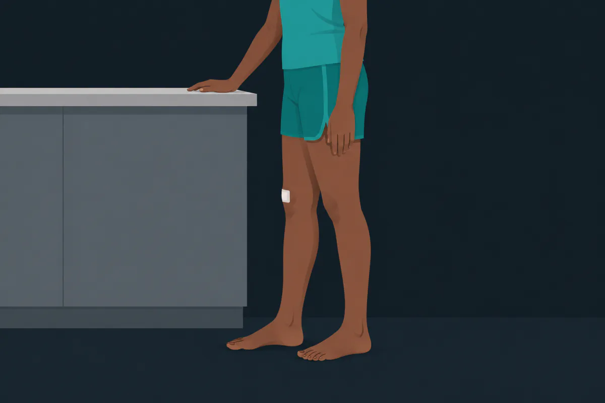
None of those is specific. Extension can be limited by swelling, by pain, by hamstring spasm, by graft placement or by scar tissue, and telling them apart is a clinical job. One distinction is worth knowing, though, because it changes what is being discussed: an extension lag, where the knee straightens fully when someone else moves it but sags when you hold it up yourself, is a muscle problem rather than a mechanical block. A cyclops lesion blocks the joint in both. The wider account of extension loss and its causes is in why won’t my knee straighten.
Why extension gets watched so closely
Full extension early is the one thing every ACL protocol agrees on, for reasons set out in Noyes’ Knee Disorders and in the consensus guideline from van Melick and colleagues, which puts range of motion ahead of loading in the order of business. Cyclops syndrome is part of the reason.
Delaloye and colleagues’ analysis of those 3,633 reconstructions found that a knee extension deficit in the early postoperative period predisposed patients to cyclops syndrome — the finding is in their title. Cause and effect are genuinely tangled here: a knee that will not straighten may be developing a nodule, and a knee that is not being straightened may be giving one room to organise. Either way, the practical consequence is the same, and it is why physiotherapists keep measuring extension rather than waiting to see. Meissburger and colleagues found the pattern again after treatment: among patients operated on for a cyclops lesion, those left with an extension deficit six weeks later were more likely to have it come back.
Separately, a knee that locks and will not move at all is one of the warning signs the NHS lists after this surgery, alongside a calf that is hot, red, swollen or painful; a fever with spreading redness or discharge at the incision; and new weakness or numbness in the foot. Any of those means contacting your surgical team, your GP or an urgent care service the same day.
What is usually done about it
First, the ordinary things: more attention to extension in rehabilitation, and time. Many extension deficits settle without surgery, which is why an unresolving one is what gets investigated rather than a stiff fortnight.
When a nodule is confirmed and the block persists, the treatment is an arthroscopic clear-out — arthrolysis, keyhole removal of the tissue. Jackson and Schaefer reported that extension improved in all of their cases after debridement. Tarchichi and colleagues, reporting on 197 of these operations, found 92 per cent of patients would have the procedure again, with a mean score of 80 on the IKDC questionnaire, a standard measure of knee function out of 100, and no significant difference between earlier and later debridement for return to sport or function. Recurrence happens but is uncommon: Meissburger and colleagues put it at 6.9 per cent after a first arthrolysis.
Where the app fits
Extension is the number to keep a record of, because a plateau is much easier to see in a column of figures than in memory — and “it has not changed in three weeks” is a more useful thing to tell a physiotherapist than “it feels stuck”. In ACL Recovery Tracker, a free ACL rehab app for iPhone and Android, the mobility log stores extension and flexion in degrees, typed in or measured with your phone’s gyroscope, with your healthy leg’s baseline to compare against; full extension is written into the progression criteria for Levels 2 and 3, so a level you cannot complete is itself a signal worth raising.
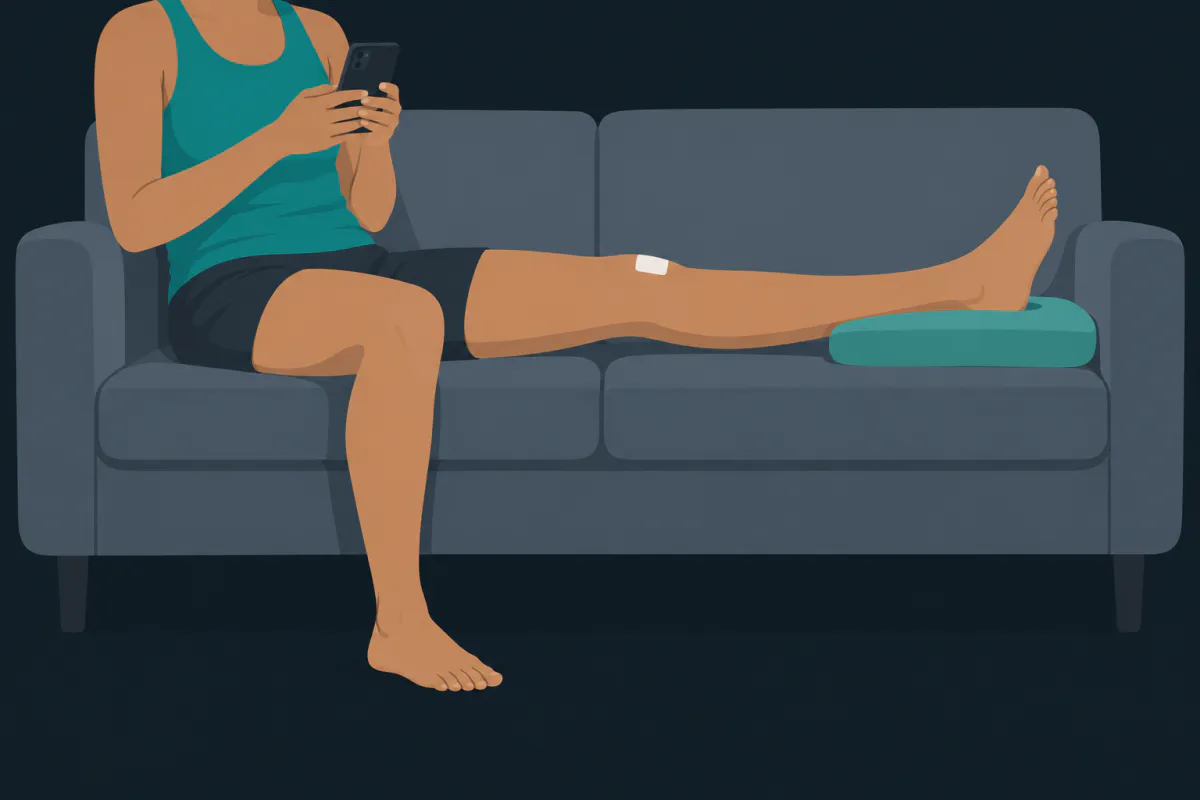
A cyclops lesion is an unglamorous problem with a memorable name, and the name does most of the damage. On the evidence, the tissue is common, the trouble is not, and when it does cause trouble the fix is a short arthroscopic procedure that patients in the largest published series were glad to have had. What that leaves worth doing is the dull part: measuring the last few degrees, and saying so when they stop moving.
The illustrations in this article were generated with AI.
Sources
- Jackson DW, Schaefer RK. Cyclops syndrome: loss of extension following intra-articular anterior cruciate ligament reconstruction. Arthroscopy. 1990;6(3):171-178.
- Noailles et al. Incidence and risk factors for cyclops syndrome after anterior cruciate ligament reconstruction: a systematic literature review. Orthop Traumatol Surg Res. 2019;105(7):1401-1405.
- Facchetti L, Schwaiger BJ, Gersing AS, et al. Cyclops lesions detected by MRI are frequent findings after ACL surgical reconstruction but do not impact clinical outcome over 2 years. Eur Radiol. 2017;27(8):3499-3508.
- Delaloye J, Murar J, Vieira TD, et al. Knee extension deficit in the early postoperative period predisposes to cyclops syndrome after anterior cruciate ligament reconstruction: a risk factor analysis in 3633 patients from the SANTI Study Group database. Am J Sports Med. 2020;48(3).
- Tarchichi et al. Good satisfaction and functional outcomes after arthroscopic debridement of cyclops syndrome post-anterior cruciate ligament reconstruction: analysis of 197 patients of the MERIscience cohort. Knee Surg Sports Traumatol Arthrosc. 2025.
- Meissburger V, Lefèvre N, Moussa MK, et al. Recurrence of cyclops syndrome after initial arthrolysis: characteristics and risk factors after ACL reconstruction. Orthop J Sports Med. 2025.
- Aloisi G, Goderecci R, Fidanza A, Cipolloni G, Calvisi V. Histological and clinical analysis of knee cyclops lesions. Acta Biomed. 2023;94(3):e2023125.
- van Melick N, et al. Evidence-based clinical practice update: practice guidelines for anterior cruciate ligament rehabilitation based on a systematic review and multidisciplinary consensus. Br J Sports Med. 2016;50:1506-1515.
- Noyes FR, Barber-Westin SD, eds. Noyes' Knee Disorders: Surgery, Rehabilitation, Clinical Outcomes. 2nd ed. Elsevier; 2017.
- NHS. ACL (anterior cruciate ligament) surgery: recovering.